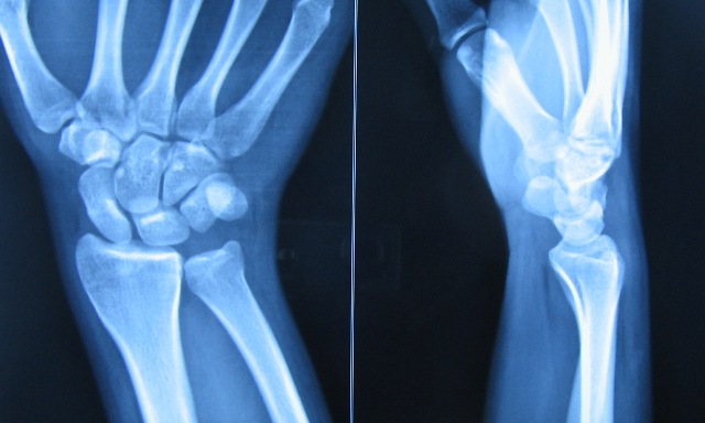

The osseous anatomy of the wrist includes the distal radius and ulna, eight carpal bones, and the five metacarpals (Figure 1). The purpose of the article is to provide insight into the various imaging modalities as they relate to imaging of the wrist in diagnosing pain and dysfunction from different types of pathology. We are now fortunate to have advanced imaging techniques to assess and diagnose the pathology of this complicated anatomic structure.

Perhaps it is no coincidence that the first human radiograph taken in 1895 by William Roentgen was that of the hand and wrist, though far from diagnostic in such primitive form. Trauma or other pathology, including degeneration, to any of these structures, can cause a wide range of pain and dysfunction that may not be easily diagnosed due to the inherent anatomic complexity. The radial and ulnar arteries, as well as their terminal branches, provide vascularity to the hand and wrist while the median, ulnar, and radial nerves are responsible for motor and sensory innervation. As with other joints, numerous ligaments and tendons connect and provide structural stability and movement of the osseous structures of the hand and wrist. Made up of eight carpal bones, the radius, ulna, and five metacarpals, the number of articulations and degrees of motion are quite extensive. Categorically considered a hinge joint like the elbow, the wrist has additional planes of movement and rotation thanks to robust anatomy. The wrist is one of the most complex joints in the human body, allowing for stability during movement in all three cardinal planes of the human body.


 0 kommentar(er)
